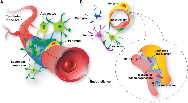
Figure 1.
Structural and molecular blood–brain barrier pericyte connections within the neurovascular unit. (A) Blood–brain barrier pericytes (yellow) and endothelial cells (red) share the basement membrane (sky blue) or are in direct contacts. In addition, blood–brain barrier pericytes are surrounded by glial cells (astrocytes, green; microglia, dark blue) and neurons (purple). (B) Blood–brain barrier pericytes and endothelial cells communicate with each other by direct contact (gap and adherens junctions) or through signalling pathways, such as platelet-derived growth factor BB (PDGF-BB)/PDGFRβ and transforming growth factor-β (TGF-β)/type 2 TGF-β receptor (TGFβR2).

