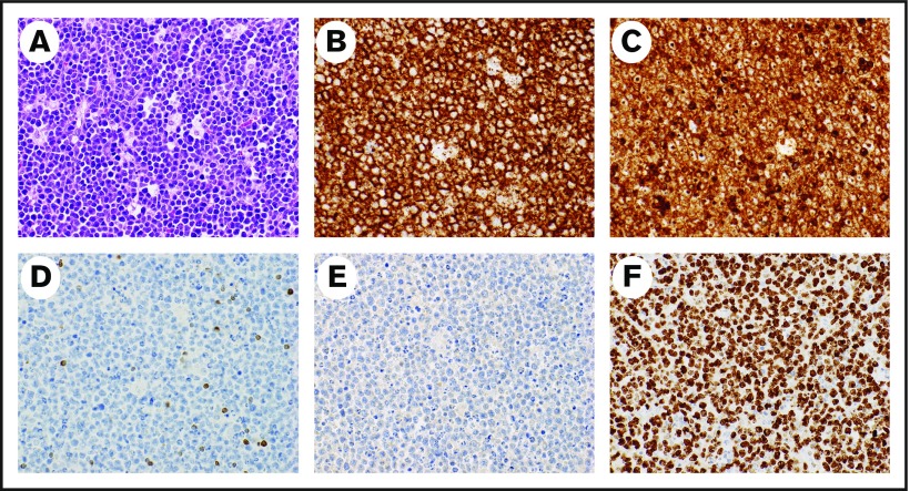
Figure 1.
Representative pathology from a patient with BL. Hematoxylin and eosin stain demonstrates typical starry-sky appearance with a diffuse infiltrate of medium-sized lymphocytes and tingible-body macrophages (A), CD20 stains the malignant cells (B), CD10 stains the malignant cells (C), BCL2 is negative in the malignant cells and stains positive in background T lymphocytes (D), TdT is negative (E), and Ki67 is positive (F) in >95% of malignant cells. Magnification ×40 for all images.

