Figure 3.
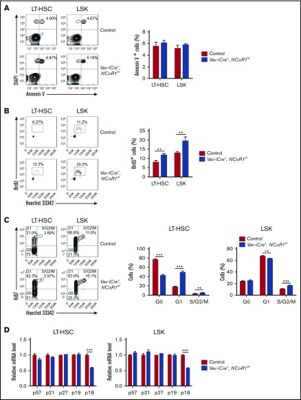
NCoR1 deficiency promotes HSC proliferation. (A) Apoptosis analysis of LT-HSCs and LSK cells in control and Vav-iCre+, NCoR1f/f mice (n = 6 per genotype). Representative FACS profiles (left panel) and the frequency of Annexin V+ cells (right panel) are shown. (B) Cell cycle analysis of LT-HSCs and LSK cells in control and Vav-iCre+, NCoR1f/f mice (n = 5 per genotype). Representative FACS profiles (left panel) and the frequency of BrdU+ cells (right panel) are shown. (C) Cell cycle analysis of LT-HSCs and LSK cells in control and Vav-iCre+, NCoR1f/f mice (n = 5 per genotype). Representative FACS profiles (left panel) and the frequency of cell cycle distribution (left and middle panels) are shown. (D) Real-time qPCR analysis of the expression of cycling-dependent kinase inhibitors genes (n = 3). mRNA levels were normalized to Gapdh expression. Data are mean ± standard error of the mean from 3 independent experiments. **P < .01, ***P < .001.

