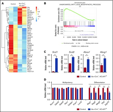
Figure 5.
NCoR1 deletion changes the gene-expression profile. (A) Heat map depicting a portion of significantly dysregulated genes in Vav-iCre+, NCoR1f/f LSK cells compared with control LSK cells (fold change > 2; false discovery rate < 0.05). See also supplemental Table 1. (B) Gene set enrichment analysis of control and Vav-iCre+, NCoR1f/f LSK cells. (C) Relative expression levels of Scd1, Scd2, Abca1, and Abcg1 in control and Vav-iCre+, NCoR1f/f LSK cells, as measured by real-time PCR (n = 3). mRNA levels were normalized to Gapdh expression. (D) Relative expression levels of select multipotency and differentiation genes in control and Vav-iCre+, NCoR1f/f LSK cells measured by real-time PCR analysis (n = 3). mRNA levels were normalized to Gapdh expression. *P < .05, **P < .01, ***P < .001. FWER, family wise error rate; NES, normalized enrichment score.

