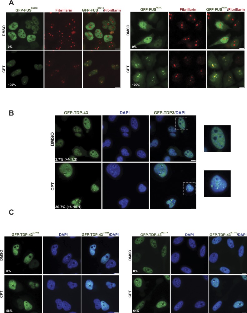
Figure S1. GFP-tagged FUS and GFP-TDP-43 relocalisation to nucleoli is not affected in HeLa cells by ALS-associated mutations, following CPT treatment.
(A) The GFP-tagged ALS-related mutant proteins GFP-FUSR521C (left) and GFP-FUSP525L (right) were detected by direct fluorescence microscopy in HeLa cells. The cells were treated with DMSO vehicle or 4 μm CPT for 1 h. Anti-fibrillarin immunostaining was used to stain nucleoli. Numbers are the percentage of GFP-positive cells with GFP-R521C or GFP-P525L nucleolar localization (n = 100 cells). Scale bars, 10 μm. (B) GFP-TDP-43 fluorescence was detected in HeLa cells treated as above. Zoomed areas are shown (right). Numbers are the mean (±SD) of the percentage of GFP-positive cells with GFP-TDP-43 nucleolar localization from three independent experiments (n = 50 cells/experiment). Scale bars, 10 μm. (C) GFP fluorescence was detected as above in HeLa cells expressing the ALS-related mutant proteins GFP-TDP-43G298S (left) or GFP-TDP-43M337V (right). Numbers are the percentage of GFP-positive cells with GFP-G298S or GFP-M337V nucleolar localization (n = 100 cells). Scale bars, 10 μm.

