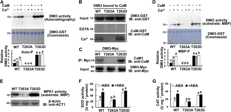
Figure 8.
Thr-263 Autophosphorylation Is Necessary for the Activation of DMI3 and the Response It Mediates.
(A) Autophosphorylation of DMI3, DMI3T263A, and DMI3T263D in vitro. Autophosphorylation activity in the presence of either 5 mM EGTA (−), 0.1 mM CaCl2, or 0.1 mM CaCl2 and 1 μM CaM was analyzed by in vitro kinase assay (top) and quantified by Quantity One software (bottom; the activity of DMI3 wild type [WT] without any treatment was set to 1). Corresponding Coomassie staining was also shown (middle).
(B) CaM binding of DMI3, DMI3T263A, and DMI3T263D in vitro. The CaM binding affinities of the mutant and wild-type protein extracts were tested in the presence of 1 mM CaCl2 (bottom) or 5 mM EGTA (middle).
(C) CaM binding of DMI3, DMI3T263A, and DMI3T263D in rice protoplasts. The indicated constructs were transfected into rice protoplasts. Total isolated proteins were incubated with an anti-Myc antibody and were detected by IB using an anti-CaM antibody (top). The input protein is shown by IB analysis using an anti-Myc antibody (bottom).
(D) Substrate phosphorylation of DMI3, DMI3T263A, and DMI3T263D in vitro. Images show substrate phosphorylation (top, MBP as substrate), the corresponding Coomassie staining (middle), and the relative activity of substrate phosphorylation (bottom). The activity of untreated DMI3 wild type was set to 1.
(E) The activity of MPK1 in the protoplasts with transiently expressed DMI3 or DMI3T263A or DMI3T263D. The activity of MPK1 in the protoplasts was analyzed by immunoprecipitation kinase assay (top). β-actin was used as a loading control (bottom).
(F) and (G) The activities of SOD (F) and CAT (G) in protoplasts transiently expressing DMI3 or DMI3T263A or DMI3T263D. Protoplasts were treated with 10 μM ABA for 10 min. All experiments were repeated at least three times with similar results. In (A), (D), (F), and (G), values are means ± sem of three independent experiments. Means denoted by the same letter did not significantly differ at P < 0.05 according to Duncan’s multiple range test. Molecular mass markers in kilodaltons were shown on the left.

