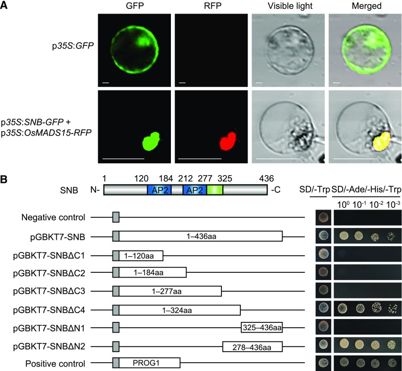
Figure 4.
Subcellular Localization and Transcription Activity of SNB.
(A) Subcellular localization of the SNB-GFP fusion protein in a rice protoplast. The OsMADS15-RFP fusion protein was used as a nuclear localization marker, and the GFP protein alone was used as the control. Bars = 20 μm.
(B) Transcription activity assay of full-length or truncated SNB in yeast. pGBKT7-SNB, pGBKT7-SNBΔC (1–4), and pGBKT7-SNBΔN (1 and 2) had the GAL4 BD (gray) in the pGBKT7 vector fused with sequences encoding full-length, N-terminal, or C-terminal SNB, respectively. pGBKT7 was used as the negative control, and the transcription factor PROSTRATE GROWTH1 was fused with the GAL4 BD as the positive control. The numbers in the boxes indicate the SNB amino acid residues used for construction.

