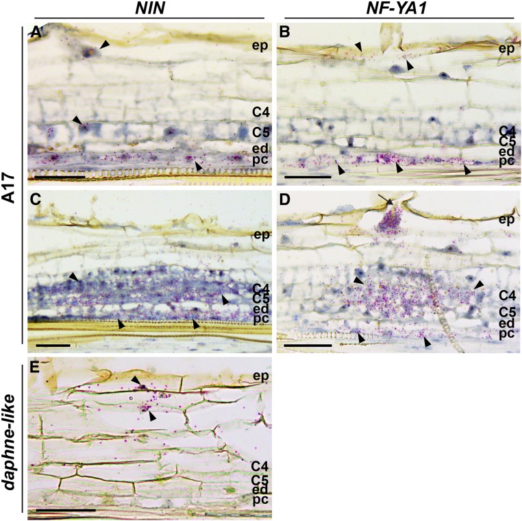
Figure 7.
NIN and NF-YA1 Expression Patterns in Medicago Wild-Type (A17) Nodule Primordia and daphne-like Mutant.
(A) to (D) RNA in situ localization of NIN ([A] and [C]) and NF-YA1 ([B] and [D]) in nodule primordia at 2 dpi ([A] and [B]) and at 3 dpi ([C] and [D]). The arrow indicates an infection thread.
(E) RNA in situ localization of NIN in roots of the daphne-like mutant at 2 dpi.
Hybridization signals are visible as red dots (arrowheads). C4 and C5, cortical cell layers 4 and 5; ed, endodermis; ep, epidermis; pc, pericycle. Bars = 50 µm.

