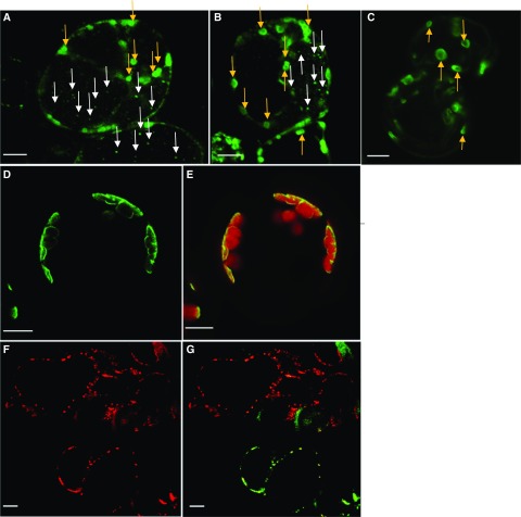
Figure 3.
Subcellular Localization of PAPST2 Observed by Confocal Fluorescence Microscopy.
(A) and (B) Transient expression of the PAPST2-GFP fusion protein under the control of the 35S CaMV promoter in Arabidopsis suspension cells derived from roots (Berger et al., 2007). (A) and (B) show two independent experiments performed using the same cell culture. The green fluorescence labels plastids (green dots— indicated with yellow arrows) and mitochondria (tiny dots—indicated with white arrows).
(C) Localization of triosephosphate/phosphate translocator -GFP—a positive control for plastids (Gigolashvili et al., 2009, 2012).
(D) and (E) PAPST2-GFP under the control of the 35S CaMV promoter in Arabidopsis mesophyll protoplasts; the green fluorescence surrounds the red autofluorescence of the chloroplasts.
(F) and (G) Co-localization of mitochondrial HSP90 protein fused to mCHERRY with the PAPST2-GFP fusion protein driven by the PAPST2 promoter in Arabidopsis dark-grown suspension cells derived from mesophyll cells. (F) Red fluorescence of the mitochondrial HSP90-mCHERRY. (G) Co-expression of PAPST2-GFP driven by the PAPST2 promoter (green) with the mitochondrial HSP90-mCHERRY (red). Cells co-expressing both constructs are shown in yellow. Red, green, and yellow dots label mitochondria.
(A) to (G) Bars = 10 µm.

