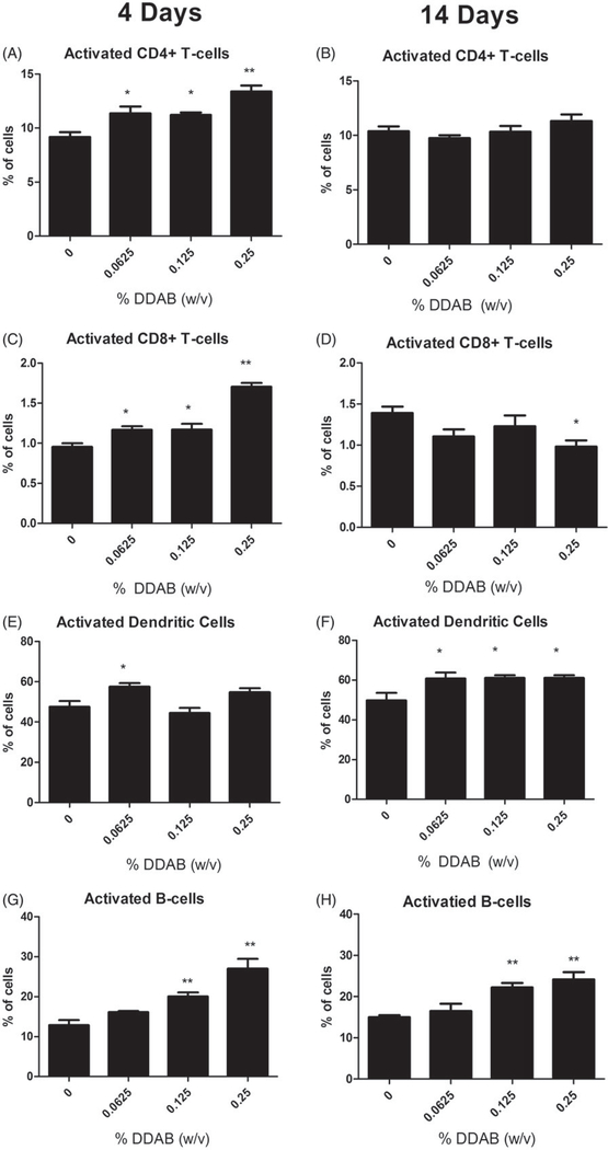
Figure 3.
Increases in activated leukocytes following dermal exposure to DDAB. Analysis of percent CD44-high CD4+ (A), CD44-high CD8+ (C), CD86+ dendritic cells (E), and CD86+ B-cells (G) of total lymphocytes on Day 10 following four days of DDAC application. Analysis of percent CD44-high CD4+ (B), CD44-high CD8+ (D), CD86+ dendritic cells (F), and CD86+ B-cells (H) of total lymphocytes on Day 15 following 14 days of DDAC application. Bars represent mean (±SE) of five mice per group. Significantly different from acetone controls at *p < 0.05 or **p < 0.01.

