Fig. 5.
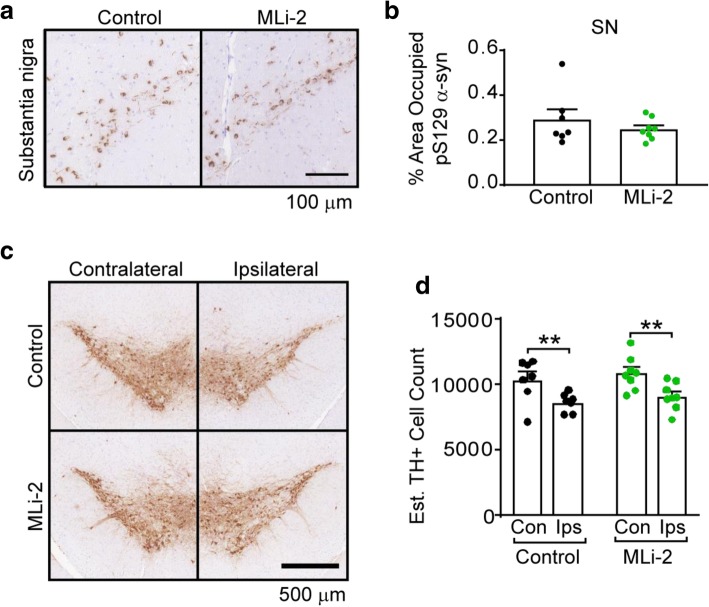
MLi-2 treatment does not impart protection from α-synuclein pathology or neuron death. a Both control and MLi-2 treated mice accumulate pS129 α-synuclein aggregates in the substantia nigra ipsilateral to the site of injection. Scale bars = 100 μm. b Quantification of pS129 α-synuclein pathology in every 10th section through the ipsilateral substantia nigra reveals that there is no difference between pathological burden in control and MLi-2 mice (p = 0.3995, unpaired t test with Welch’s correction). c The accumulation of α-synuclein pathology leads to substantial loss of substantia nigra neurons in both control and MLi-2 treated mice ipsilateral to the site of injection. This is shown here through use of TH staining. Scale bar = 500 μm. d Every 10th section through the midbrain was stained with TH and TH-positive cells were counted to estimate the total number of neurons in the substantia nigra. While there was significant loss of neurons ipsilateral to the site of injection (control p = 0.0048, MLi-2 p = 0.0026, two-way ANOVA followed by Sidak’s multiple comparisons test), there was no significant difference between control and MLi-2 treated mice (control versus MLi-2 ipsilateral to injection p = 0.6988, two-way ANOVA followed by Sidak’s multiple comparisons test). All plots are means and error bars represent s.e.m. with individual values plotted

