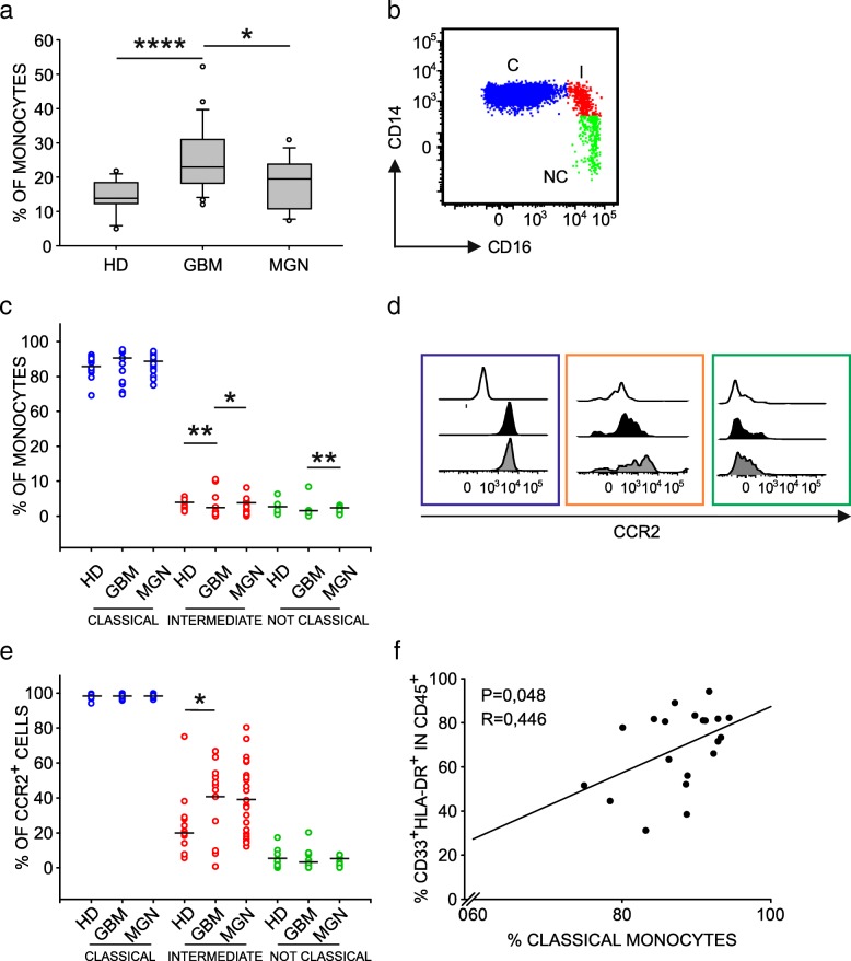
Fig. 6.
Characterization of monocyte dysregulation in GBM patients. a Boxplots of the distribution of percentage of monocytes in blood samples from HD (n = 12), GBM (n = 24) and meningioma (MNG) (n = 13) patients, calculated as HLA-DR+ cells among PBMCs. b Analysis of monocyte subsets in whole blood using CD14 and CD16 markers. Dot plot gated on PBMCs shows classical monocytes (C) CD14high/ CD16− and intermediate subset (I) and a non-classical subset (NC). (c) Distribution of C, I and NC monocytes in meningioma (MNG, n = 13) and glioblastoma patients (GBM, n = 24) in comparison to HDs (n = 12). d Representative example of CCR2 expression on monocyte subsets in GBM patients. White histograms show fluorescence minus one (FMO) controls, black histograms refer to MFI values of CCR2+ cells of healthy donor, while grey histograms indicate MFI values of CCR2+ cells among C (blue), I (orange), and NC (green) monocytes of GBM patients. e Cumulative data showing CCR2 expression in MNG (n = 13) and GBM patients (n = 24) compared to HDs (n = 12). Mann-Whitney test for statistical significance between pairwise groups. f Correlation between the percentage of classical monocytes in PBMCs and that of macrophages among GBM-infiltrating leukocytes. Spearman’s rank-order correlation on 20 paired samples

