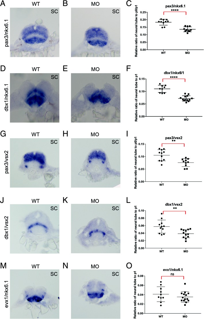
Fig. 8.
prdm12b controls the size of the p1 domain. Expression of pax3/nkx6.1 (a, b), dbx1/nkx6.1 (d, e), pax3/vsx2 (g, h), dbx1/vsx2 (j, k) and evx1/nkx6.1 (m, n) in 24hpf wildtype (a, d, g, j, m) or prdm12b MO-injected (b, e, h, k, n) embryos. Panels show cross sections through the spinal cord with dorsal to the top. c, f, i, l, o show quantification of the size (along the dorsoventral axis) of the p0/p1 domain (c, i) or the p1 domain (f, l, o) relative to the neural tube. At least 10 representative sections were used for each gene pair

