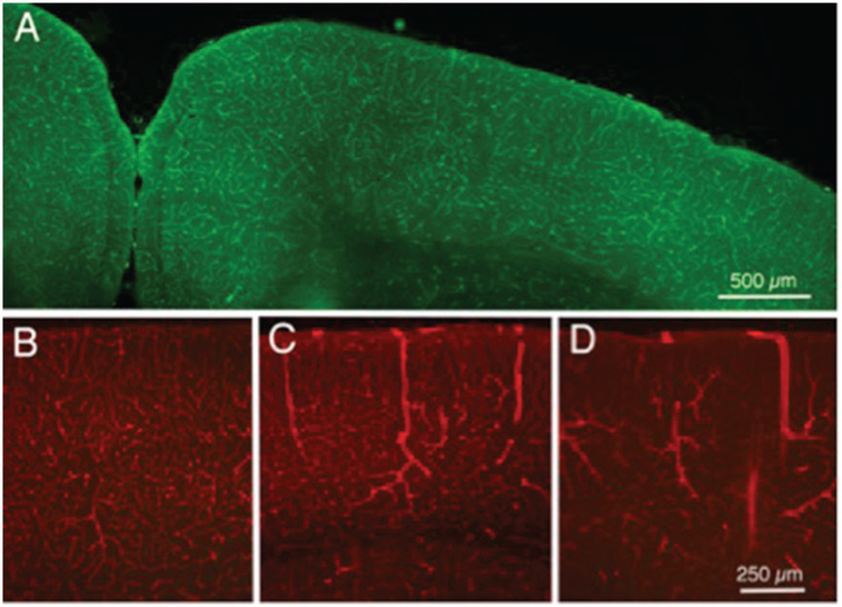
Figure 2.
Fluorescence photomicrographs of sections of cerebral cortex from mice that received intravenous injections of labeled tomato lectin. (a) Normal control mouse, injected with labeled 488 nm lectin. (b) Another normal control mouse, injected with 594 (red) labeled lectin; most labeled vessels are likely capillaries, although arrows indicate larger vessels, likely venules. (c) Mouse injected with 594 lectin 24 h following a single TBI head impact of moderate strength; note the three dilated vessels in cerebral cortex. (d) Mouse injected with 594 lectin after five daily impacts of moderate strength; note the several dilated vessels in cortex (arrows), as well as the reduced numbers of normal appearing capillaries.

