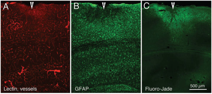
Figure 5.
Fluorescence photomicrographs of three adjacent sections from mouse cerebral cortex demonstrating the effects of a series of five impacts (moderate strength), seven days after the last impact. (a) Cortical vasculature, as indicated by 594 (red) labeled lectin. (b) Astrocyte proliferation, as indicated by 488 (green) labeling for GFAP immunohistochemistry. (c) Neuronal degeneration, as indicated by Fluoro-Jade labeling. Arrowheads indicate site of cortical damage following the TBIs. Calibration bar in C = 500 μm for all images.

