
Figure 5.
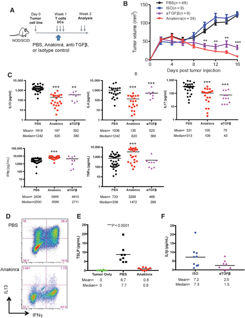
IL1 and TGFβ mediate tumor-promoting type 2 cytokines in humanized mouse model. Hs578T breast tumor-bearing NOD/SCIDβ2–/– mice were reconstituted with MDDCs and autologous T cells. Mice were treated with either: (i) anti-TGFβ neutralizing antibody on days 3, 6, and 9; (ii) anakinra daily starting on day 3; or (iii) controls such as isotype and PBS. A, Scheme of experimental mouse model. B, Kinetics of tumor growth from multiple experiments. Number of mice in each group is indicated. C, Breast tumor fragments were harvested at day 16 after tumor implantation and stimulated for 16 hours with PMA and ionomycin. Cytokines were measured by Luminex. D, Cell suspensions stained for IL13 and IFNγ expression by FACS. Representative plots from three different mice. E, TSLP concentration by Luminex in tumor only versus PBS control versus anakinra group. F, IL1β concentration by Luminex in isotype control versus anti-TGFb neutralizing antibody group.