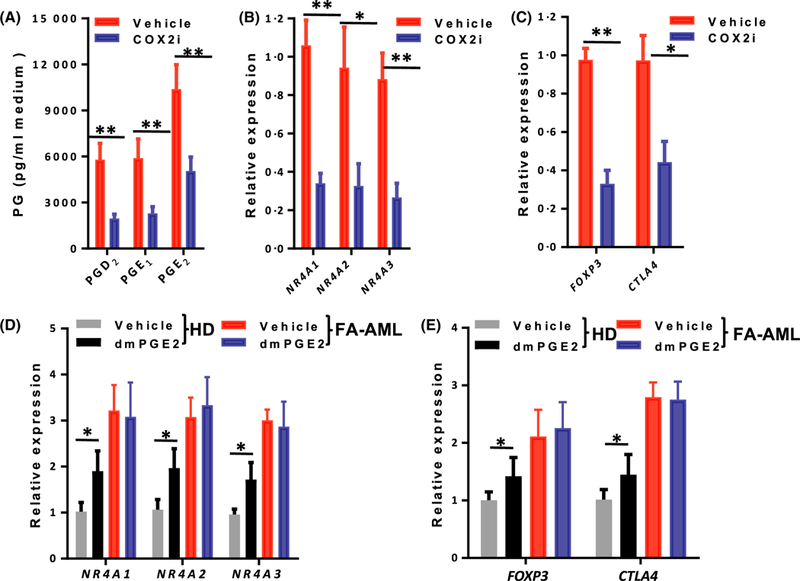
Fig 4.

The link between AML-MSC COX2-PG secretome and NR4A-Treg signalling. (A) Mesenchymal inhibition of COX2 reduces the secretion of prostaglandins (PGs). Mesenchymal stromal cells (MSCs) from Fanconi anaemia (FA) patients with acute myeloid leukaemia (FA-AML) were pre-treated with COX2 inhibitor (celecoxib, 10 μmol/l) or vehicle (5% dimethyl sulphoxide) for 2 h, and the levels of the indicated PGs in the culture medium were measured by enzyme-linked immunosorbent assay. Results are means ± SD of three independent experiments (n = 9 for each group). (B) Mesenchymal inhibition of COX2 reduces the expression of the NR4A transcription factors (TFs) in co-cultured CD34+ cells. The MSCs in (A) were co-cultured with normal CD34+ cells for 2 weeks and expression of the three NR4A TF genes was determined by real-time polymerase chain reaction (PCR). Results are means ± SD of three independent experiments (n = 9 for each group). (C) Mesenchymal inhibition of COX2 reduces the expression of Treg genes in co-cultured CD34+ cells. The MSCs in (A) were co-cultured with normal CD34+ cells for 2 weeks and expression of FOXP3 and CTLA4 was determined by real-time PCR. The normal CD34+ cells were from 12 healthy donors. Results are means ± SD of three independent experiments (n = 9 for each group). (D) 16–16 dimethyl-PGE2 (dmPGE2) treatment increases the expression of the NR4A TF genes in co-cultured CD34+ cells. MSCs derived from HD were pre-treated with dmPGE2 followed by co-culture with normal CD34+ cells for 2 weeks. The expression of the three NR4A TF genes was determined by real-time PCR. Results are means ± SD of three independent experiments (n = 9 for each group). (E) dmPGE2 treatment increases the expression of Treg genes in co-cultured CD34+ cells. Co-cultured normal CD34+ cells described in (D) were subjected to real-time PCR analysis for FOXP3 and CTLA4. The normal CD34+ cells were from 12 healthy donors. Samples were normalized to the level of GAPDH mRNA. Results are means ± SD of three independent experiments (n = 9 for each group). *P < 0·05, **P < 0·01. [Colour figure can be viewed at wileyonlinelibrary.com]
