Fig. 3.
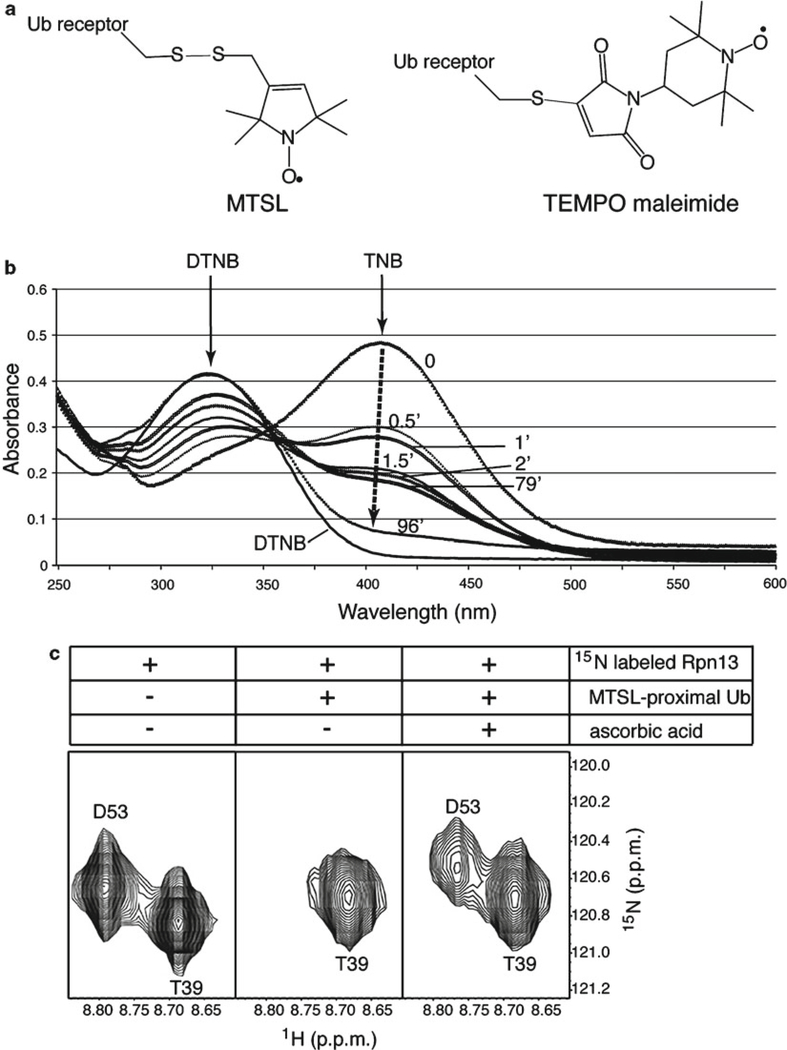
Use of spin labeling to characterize ubiquitin:receptor complexes. (a) Illustration of MTSL (left) or TEMPO maleimide (right) attached to a ubiquitin receptor by a cysteine thiol group via sulfur–sulfur or by carbon–sulfur bonds (gray), respectively. (b) Spin labeling reaction time course as measured by a DTNB assay. DTNB absorbs at 324 nm and reacts with free thiol groups to release 2-nitro-5-thiobenzoate (TNB), which absorbs at 412 nm and is not observed when spin labeling is complete (black dashed arrow). The plotted absorbance curves were recorded before adding MTSL (0 min) and after reacting it at equimolar quantity and 4°C in the dark for 0.5, 1, 1.5, 2, 79, and 96 min. (c) Expanded view of 1H, 15N HSQC spectra of hRpn13 (1–150) alone (left panel), with MTSL-labeled K48-linked diubiquitin G75C (middle panel), and with ascorbic acid added to the mixture (right panel). G75C-MTSL is on the proximal subunit and D53, but not T39, of hRpn13 experiences paramagnetic relaxation enhancement (middle), which is recovered by quenching (right) (Panel (c) is reprinted from ref. 39 with permission from Elsevier).

