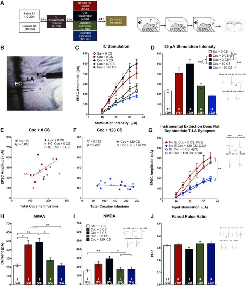
Figure 2. Cocaine SA and Cocaine-Cue Re-exposure Bidirectionally Modulate T-LA Synapses.

(A) Left: experimental timeline for electrophysiological recording-electrical stimulation experiments. Right: cartoon demonstrating the contingencies of the four experimental phases: SA, IE, cue re-exposure, and ex vivo electrophysiology.
(B) Image of an LA coronal section. EPSCs were evoked from LA principal neurons by stimulating the internal capsule (IC) (putative T-LA synapses). EC, external capsule.
(C) Input-output relationship for each group plotting average EPSC amplitude versus stimulation intensity. Cocaine SA increases EPSC amplitude relative to saline SA. Brief cue re-exposure (3 CSs) does not further alter EPSC amplitude, but moderate (60 CSs) and extended (120 CSs) cue extinction reverses cocaine-cue-induced potentiation. Two-way ANOVA, main effect of group (F(4,40) = 13.54, p < 0.001) and a stimulation (stim.) intensity x group interaction (F(20,200) = 8.96, p < 0.001).
(D) Bar graph showing average EPSC amplitude at 35 μA stimulation intensity. Post hoc analysis from (C): *p < 0.05, ***p < 0.001.
(E) EPSC amplitude (35 μA stimulation intensity) tends to be positively correlated with the number of cocaine infusions received during acquisition for cocaine-SA animals that did not receive cue re-exposure. The correlation includes all animals that were never re-exposed to the CS, including home cage (HC) and IE controls. r(18) = 0.434, #p = 0.056, n = 20.
(F) EPSC amplitude was not correlated with the number of cocaine infusions for animals that self-administered cocaine but received extensive cue extinction. The correlation includes all animals that received 120 CS re-exposure, including IE controls; r(5) = −0.089, p = 0.850, n = 7.
(G) IE does not alter T-LA synaptic strength. The EPSC input-output relationship was significantly higher for rats that received no cue re-exposure (0 CSs) compared with rats that received extended extinction (120 CSs), independent of IE. Two-way ANOVA, main effect of group (F(3,24) = 8.38, p < 0.001) and a stimulation intensity x group interaction (F(15,120) = 4.87, p < 0.001); post hoc analysis: **p < 0.01, ***p < 0.001.
(H) Cocaine-cue memory manipulations significantly affect AMPAR current (one-way ANOVA, F(4,34) = 12.70, p < 0.001); post hoc analysis: *p < 0.05, **p < 0.01.
(I) Cocaine-cue memory manipulations significantly affect NMDAR current (one-way ANOVA, F(4,34) = 5.77, p = 0.001); post hoc analysis: *p < 0.05, **p < 0.01.
(J) Cocaine-cue memory manipulations do not alter PPR (one-way ANOVA, F(4,40) = 1.81, p = 0.140). Insets, sample average EPSC traces evoked at reverse potential (Erev) −70 mV (AMPA, PPR) and Erev +40 mV (NMDA); scale bars, 50 ms, 200 pA; error bars, mean ± SEM, n in italics = number of rats (number of neurons). See also Figures S1–S4.
