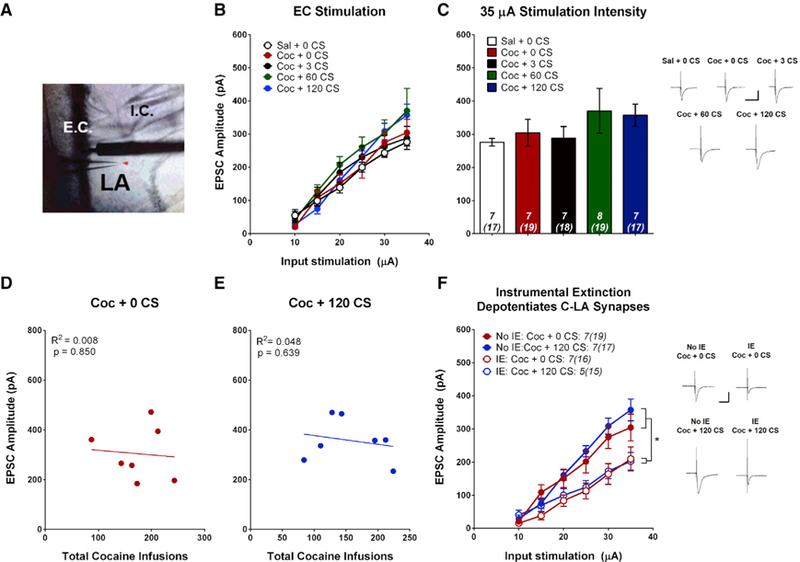
Figure 3. C-LA Synapses Are Altered by IE but Not by Cocaine-Cue Memory Manipulations.

(A) Image of LA coronal section. EPSCs were evoked from LA principal neurons by stimulating the EC (putative C-LA synapses).
(B) Input-output relationship for each group, plotting average EPSC amplitude versus stimulation intensity. Neither cocaine SA nor cue re-exposure significantly alter EPSC amplitude at C-LA synapses (two-way ANOVA, F(4,31) = 0.854, p = 0.503).
(C) Bar graph showing average EPSC amplitude at 35 μA stimulation intensity.
(D) EPSC amplitude (35 μA stimulation intensity) was not correlated with the number of cocaine infusions during acquisition for cocaine-SA animals that did not receive cue re-exposure (r(5) = −0.0891, p = 0.850, n = 7).
(E) EPSC amplitude (35 μA stimulation intensity) was not correlated with the number of cocaine infusions earned during acquisition for cocaine-SA animals that received extensive cue extinction (r(5) = −0.219, p = 0.639, n = 7).
(F) C-LA synapses are depotentiated by IE. The EPSC input-output relationship was significantly lower for all rats that received IE, independent of cue re-exposure. Two-way ANOVA, main effect of group (F(3,22) = 5.11, p = 0.008) and a stimulation intensity x group interaction (F(15,110) = 3.69, p < 0.001); post hoc analysis: **p < 0.05.
Insets, sample average EPSC traces evoked at Erev −70 mV; scale bars, 50 ms, 200 pA; error bars, mean ± SEM; numbers in italics, number of rats (number of neurons).
