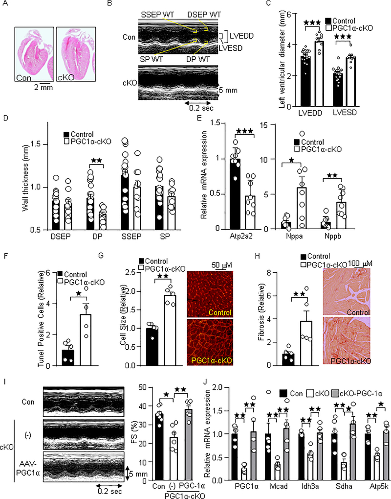
Fig. 2.
Cardiac-specific loss of PGC-1α results in heart failure. (A) H&E stained images of the PGC-1α-cKO mice. (B) Representative M-mode echocardiography of the PGC-1α-cKO mice. (C) Enlarged left ventricular diameter and (D) reduced wall thickness (WT). LVEDD: LV end diastolic dimension, LVESD: LV end systolic dimension, DSEP: Diastolic septal, DP: Distolic posterior: SP: Systolic posterior, SSEP: systolic septal. (E) Heart failure marker genes expression in PGC-1α-cKO mice. The expression of indicated genes was examined by quantitative real time PCR (N=8). Atp2a2: ATPase, Ca++ transporting, cardiac muscle, slow twitch 2, Nppa: Natriuretic peptide A, and Nppb: Natriuretic peptide B. Histological analyses of myocardium in PGC-1α-cKO mice. Histochemical analyses were performed with (F) Tunel, (G) WGA, and (H) PSR staining. (I) Re-expression of PGC-1α with AAV normalizes cardiac systolic dysfunction in PGC-1α-cKO mice. Representative M-mode echocardiography (Left). Fractional shortening (Right). (J) Re-expression of PGC-1α with AAV normalizes PGC-1α target gene expression in PGC-1α-cKO mice. Echocardiographic measurements (I) and gene expression analysis (J) were performed after 3 weeks of transduction of AAV- PGC-1α and the control (GFP). The numbers of mice examined in each experimental group were: 6–17(C-D), 8 (E), 4–5 (F-H), 5–7 (I) and 4–6 (J). * p<0.05; ** p<0.01; *** p<0.001 as indicated.

