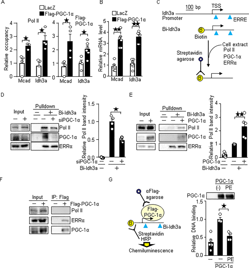
Fig. 6.
PGC-1α promotes Pol II recruitment. (A-B) Overexpression of PGC-1α promotes Pol II recruitment and target gene expression in cardiomyocytes. Flag-PGC-1α was overexpressed with adenovirus vector. (A) ChIP assays were performed with anti-Pol II and anti-Flag antibodies. N=3–8. (B) The expression levels of indicated genes were examined. (C) Schematic representation for the DNA binding assay. Biotin-labeled Idh3a promoter containing ERRE and TSS were incubated with cell lysate. The PGC-1α, ERRα and Pol II bound to the DNA was examined with Western blot analyses. (D) Knockdown of PGC-1α inhibits Pol II recruitment in vitro. N=4. (E) Overexpression of PGC-1α promotes Pol II recruitment. N=5. (F) The binding of PGC-1α to Pol II was not observed with co-immunoprecipitation assay. Flag-PGC-1α was overexpressed in cardiomyocytes with adenovirus vector. Co-immunoprecipitation assays were performed with anti-Flag antibody. (G) PGC-1α purified from phenylephrine (PE) treated cells has a lesser ability to bind to the promoter. Cardiomyocytes expressed Flag-PGC-1α were treated with 100 μM PE for 16 hours. Flag-PGC-1α was immunoprecipitated with anti-Flag-antibody. The immunocomplex was incubated with HEK293 cell lysate as a source of ERRs and general transcriptional machineries, biotin-labeled Idh3a promoter and HRP (horseradish peroxidase)-conjugated streptavidin. The binding of PGC-1α and biotin-labeled Idh3a promoter was measured by chemiluminescence. N=4–5. * p<0.05; ** p<0.01 as indicated.

