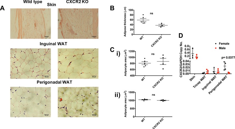
Figure 3.

CXCR2 KO male mice have no significant change in adipocyte size compared to wild‐types. (A) Skin and adipose depots were dissected from adult male mice (>8 wk) before processing and H&E staining of sections from wild‐type and CXCR2 KO mice. Brightfield microscopy was used to take images of the skin, inguinal adipose, or perigonadal adipose tissue (scale bars: 50 μm). (B) Adipose thickness and (C) individual adipocyte area for (i) inguinal and (ii) perigonadal sites were measured. (D) Quantitative real‐time PCR was used to analyze CXCR2 expression in skin or adipose tissues from male and female mice, relative to the house‐keeping gene, GAPDH. Data are plotted as mean (±sem), where each symbol represents data from an individual mouse and analyzed using an unpaired t test or Welch's t test (D), ns = not significant
