
Figure 5.
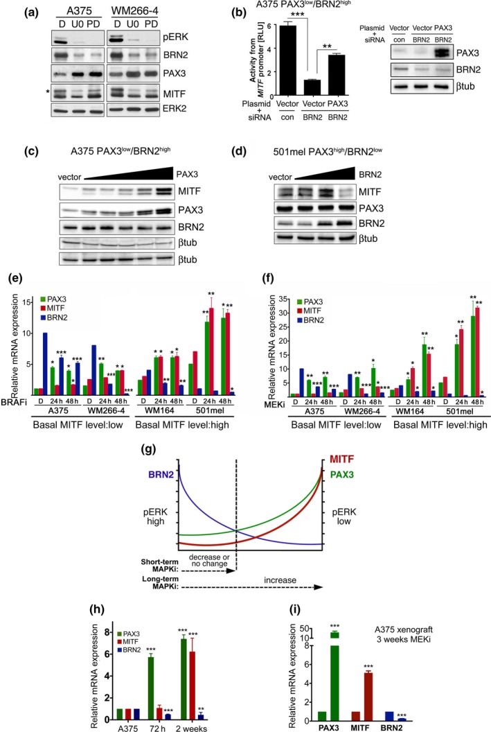
Long‐term BRAF and MEK inhibition increases PAX3 and MITF expression. (a) Western blot analysis for MITF, PAX3, BRN2, pERK and ERK2 in A375 and WM266‐4 cells treated with 10 µM U0126 (U0), 1 µM PD184352 (PD) or DMSO (D) for 24 hr. (b) Luciferase assay for the M‐MITF promoter activity in A375 cells co‐transfected with either control or BRN2 siRNAs and a PAX3 expression or control plasmid. (c) Western blot for the indicated proteins from A375 cells transfected with increasing amounts (200–600 ng) of a PAX3 expression plasmid. (d) Western blot for the indicated proteins from 501mel cells transfected with increasing amounts (300–500 ng) of a BRN2 expression plasmid. (e) RT‐qPCR analysis of PAX3, MITF, BRN2 expression in the indicated cell lines untreated or treated with 1 µM vemurafenib (BRAFi) or (f) with 1 µM AZZD6244 (MEKi). (g) Model for the regulation of MITF by PAX3 and BRN2. Short‐term MAP kinase pathway inhibition results in reduced BRN2 and increased PAX3 expression, but long‐term inhibition will lead to constant PAX3 upregulation and consequently increased MITF expression. (h) RT‐qPCR analysis of PAX3, MITF, BRN2 expression in A375 cells were treated with MEKi for the indicated times. (i) RT‐qPCR analysis for PAX3, MITF, BRN2 expression in A375 melanoma xenografts from mice treated with DMSO or with MEKi for 3 weeks. Data presented as the mean ± SEM are from at least three biological repeats