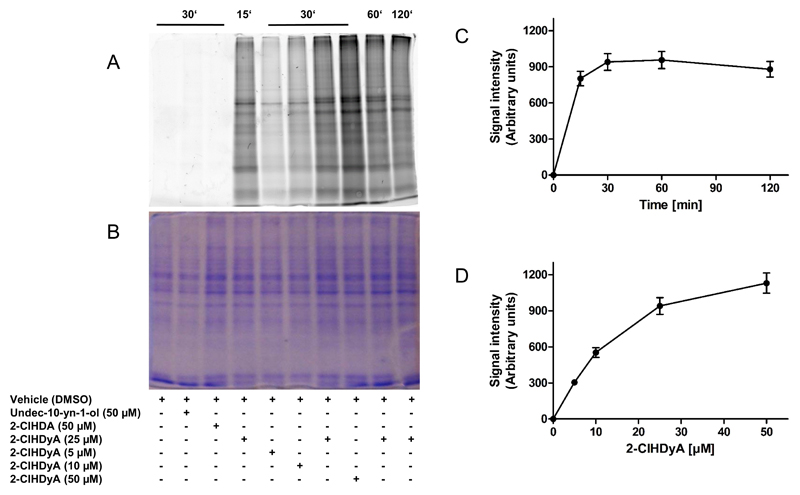
Fig. 5. 2-ClHDyA forms covalent adducts with hCMEC/D3 proteins.
Cells grown on 10 cm dishes were incubated in the absence (vehicle, 0.4% DMSO, final concentration) or presence of undec-10-yn-1-ol, 2-ClHDA, or 2-ClHDyA, followed by treatment with NaCNBH3 to reduce formed Schiff bases.
(A) Cells on 10 cm dishes were lysed in 50 mM Tris/HCl, 1% SDS, pH 8.0 and aliquots were subjected to click chemistry. After precipitation proteins were subjected to SDS-PAGE (forty μg cell protein/lane) and imaged on a Typhoon 9400 scanner.
(B) Loading control showing Coomassie Brilliant Blue stained protein lanes to verify equal loading.
(C) Time- and (D) concentration-dependent increase in fluorescence intensities of protein lanes from the image shown in (A).

