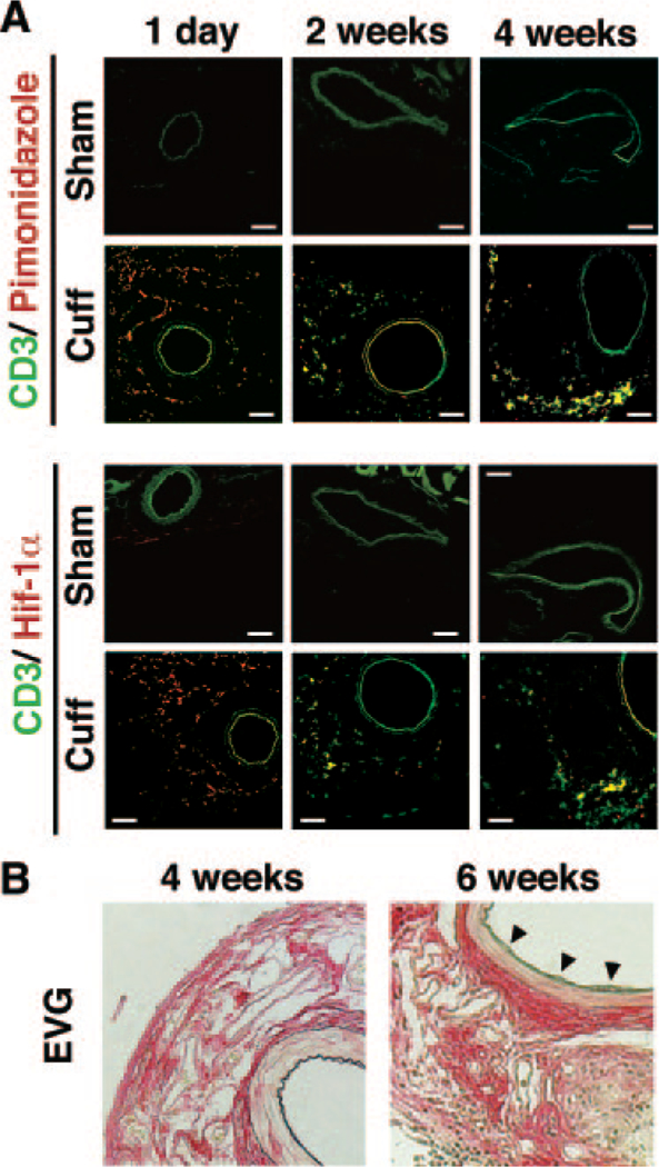
Figure 1.

Development of vascular remodeling of cuff-injured artery in mice. A, Immunofluorescence analysis of cuff-injured femoral arteries. Representative cross sections for CD3 (green), pimonidazole-protein adducts (red, upper panel), and hypoxiainducible factor (Hif) 1α (red, lower panel) at 1 and 14 days after cuff placement. Green signals of intima, media, and other muscle tissues around vessels are nonspecific signals, as proved in Supplementary Figure S1. The bar indicates 200 μm. B, Representative cross sections with hematoxylin-eosin staining of the cuffed femoral artery at 14 and 42 days after cuff placement. Elastica van Gieson staining (EVG) indicates.
