
Figure 3.
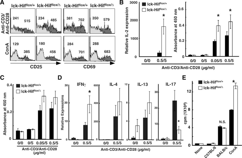
In vitro experiments on activation, proliferation, and function of Hif-1α–deficient T cells. A, Initiation of TCR signals provoking early cellular responses. After 16 hours in culture with either the combination of anti-CD3 and anti-CD28 antibodies or Con A (2.5μg/mL), lymphocytes from mandibular lymph nodes were two-color stained with phosphatidylethanolamine-labeled anti-CD4 antibody and fluorescein isothiocyanate–labeled anti-CD25 or anti-CD69 antibody or normal IgG. The profiles of CD25 and CD69 expression are shown by electrically gated CD4+ cells, in the presence of stimulants (dark gray area) or the absence of stimulants (gray area). Numbers indicate the mean fluorescence intensity of CD25 and CD69 within the cells. B, Lymphocytes from lymph nodes after 16 hours inculture with the combination of anti-CD3 and anti-CD28 antibodies were used for measurement of interleukin (IL) 2 transcripts. The culture supernatants were used for measurement of IL-2 proteins by enzyme-linked immunosorbent assay. The values are shown as absorbance at 450 nm. C, On day 2 of culture, cultured wells were tested for cell proliferation by the 3-(4,5-dimethylthiazol-2-yl)-2,5-diphenyltetrazolium bromide assay. The values are shown as absorbance at 450 nm. Values are expressed as mean_SD. *P<0.001. D, Lymphocytes from lymph nodes after 12 hours in culture with the combination of anti-CD3 and anti-CD28 antibodies were used for measurement of transcripts for interferon (IFN) γ, IL-4, IL-13, and IL-17. E, Thymidine-incorporation assay of proliferating allogeneic BALB/c lymph node lymphocytes (5.0×10⁵ cells) incubated with an equal amount of lymphocytes derived from Lck-Cre;Hif-1αflox/+ or Lck-Cre;Hif-1αflox/Δ mice. *P<0.05.