Figure 5.

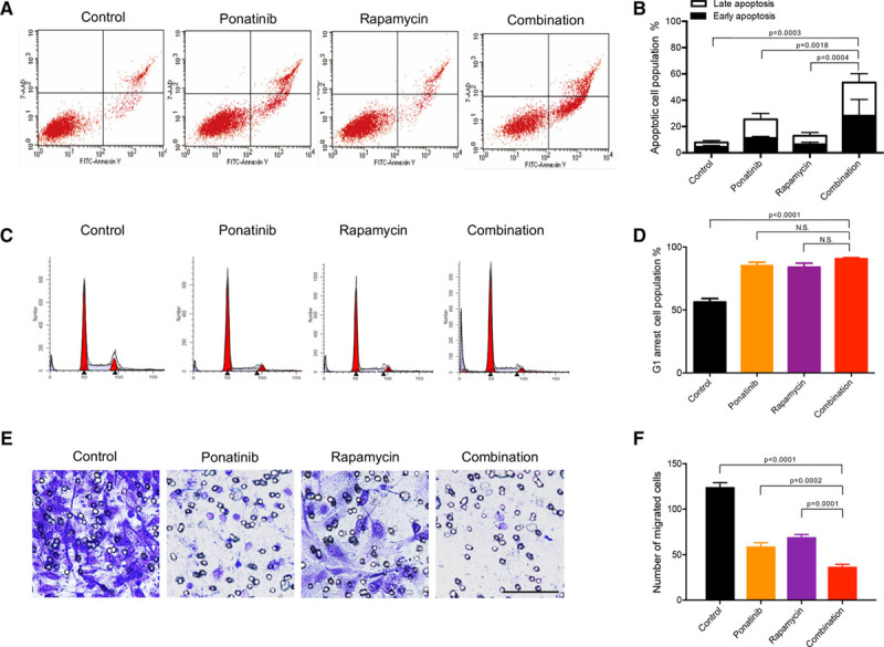
Ponatinib combined with rapamycin enhances apoptosis and inhibits cell migration in human umbilical vein endothelial cell (HUVEC)-TIE2-L914F. A, Flow cytometric analysis of apoptosis. HUVEC-TIE2-L914F cells were treated with control (dimethyl sulfoxide [DMSO]), 100 nmol/L ponatinib, 10 nmol/L rapamycin, or combination for 72 h. B, Quantification of % apoptotic cell populations. Data expressed as mean±SD, 1-way ANOVA for multiple comparisons (representative of 2 independent experiments, n=4). C, Representative images of cell cycle analysis. HUVEC-TIE2-L914F cells were treated with control (DMSO), 100 nmol/L ponatinib, 10 nmol/L rapamycin, or combination for 48 h. D, Quantification of % G0-G1 cell cycle cell population. Data expressed as mean±SD, 1-way ANOVA for multiple comparisons (representative of 2 independent experiments, n=4). E, Representative images of cells migrated through the 8 μm pore filter (Transwell migration assay). HUVEC-TIE2-L914F cells were treated with control (DMSO), 100 nmol/L ponatinib, 10 nmol/L rapamycin, or combination for 6 h and then stained with crystal violet. Scale bar: 100 μm. F, Quantification of migrated cells. Data expressed as mean±SD, 1-way ANOVA for multiple comparisons (n=3 independent experiments).
