Figure 8.

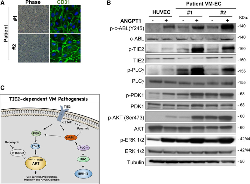
Ponatinib combined with rapamycin induces venous malformation (VM) lesion regression in a patient-derived cell xenograft model. A, Representative phase-contrast images of VM-patient-derived endothelial cell (VM-EC) cell morphology and stained with endothelial cells marker CD31 (green) and nuclei with DAPI (blue). Scale bar: phase 100 μm, IF 50 μm. B, Immunoblotting analysis of human umbilical vein endothelial cell (HUVEC), patient no. 1 and no. 2 VM-EC with indicated antibodies. Cells were treated with or without 500 nmol/L ANGPT1 (angiopoietin 1) for 15 min. C, Scheme of signaling pathways downstream of TIE2-L914F and mechanisms of action of the drug combination rapamycin+ponatinib. Rapamycin inhibited AKT (protein kinase B) activation in EC by prolonged treatment. Here, we show that ponatinib can target c-ABL but also TIE2, resulting in inhibition of AKT most likely by c-ABL effect on PI3K (phosphoinositide-3-kinase)/PDK1. Ponatinib also affected PLCγ (phospholipase C) and ERK1/2 (extracellular signal-regulated kinase), both were also decreased upon c-ABL/ARG knockdown.
