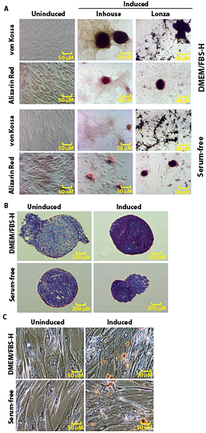
Fig 9. Trilineage differentiation of canine Ad-MSCs expanded in DMEM/FBS-H or serum-free medium containing bFGF and PDGF.

A. Osteogenic potential of canine Ad-MSCs cultured in DMEM/FBS-H or serum-free medium containing bFGF and PDGF was assessed with Alizarin Red or von Kossa staining after 14 days of induction. Canine Ad-MSCs cultured in both media types formed calcified cell aggregates. B. Chondrogenic potential of canine Ad-MSCs cultured in DMEM/FBS-H or serum-free medium containing bFGF and PDGF was assessed with Toluidine Blue staining after 14 days of induction. C. Adipogenic potential of canine Ad-MSCs cultured in DMEM/FBS-H or serum-free medium containing bFGF and PDGF was assessed with Oil Red O staining after 14 days of induction/maintenance. Uninduced cells maintained their characteristic spindle shaped morphology. Upon induction, canine Ad-MSCs cultured in both media types formed adipocytes with intracellular lipid droplets.
