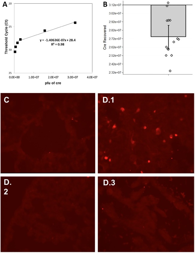
Fig 1. Validating intrathoracic injection technique.
(A) A standard calibration curve was made using known quantities of Ad5CMVCre to then quantify unknown samples of Ad5CMVCre. Six different known concentrations of Ad5CMVCre were spiked into lung tissue to make the standard calibration curve: 3.12x10⁵, 6.24x10⁵, 1.56x10⁶, 3.12x10⁶, 1.56x10⁷, and 3.12x10⁷ pfu. (B) Amount of Ad5CMVCre (in pfu) measured in 14 mice that had their left lung injected with 3.12x10⁷ pfu Ad5CMVCre virus. Measurements were made using the TaqMan real-time PCR assay. The bar plot shows the mean with standard error and overlaying dot plot shows measurements of individual animals (n = 14). pfu = plaque-forming unit, a standard measure of the number of particles capable of forming plaques per unit volume. (C-D) To characterize Ad5CMVCre activity in the left lung of KrasG12D+/-; p53fl/fl; myristolated-p110αfl/fl-ROSA-gfp mice, gfp immunohistochemistry was used to detect Cre-recombination effect within the left lung, 4 days after Ad5CMVCre injection. (C) A representative micrograph of no gfp/Cre-recombination in the left lung of a mouse that had no Ad5CMVCre virus injected serves as a negative control. (D.1) A representative micrograph of activation of gfp/Cre-recombination in the left lung of a mouse that underwent Ad5CMVCre virus injection. To validate that the virus did not enter the bloodstream to turn on other primary cancers, (D.2) is representative of no gfp/Cre-recombination activation in the right lung and (D.3) is representative of no gfp/Cre-recombination activation in the heart.

