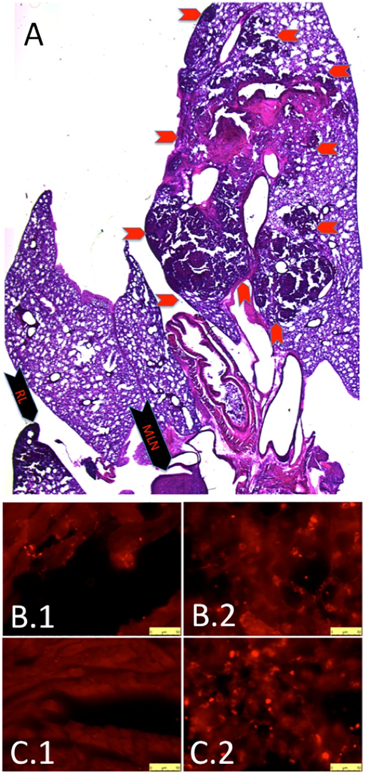
Fig 7. Metastases, histology.

(A) H&E stain confirms primary tumor in left lung (red chevrons outline border of primary tumor) as well as metastases to right lung (black chevron-RL) and mediastinal lymph nodes (black chevron-MLN). Surfactant protein C (SPC) immunohistochemistry demonstrates metastases to the heart in a CIH mouse (B.1) and Sham mouse (B.3). We provide SPC immunohistochemistry to CIH lung (B.2) and Sham lung (B.4) for positive control. All micrographs taken at 40X, bar is 50 microns.
