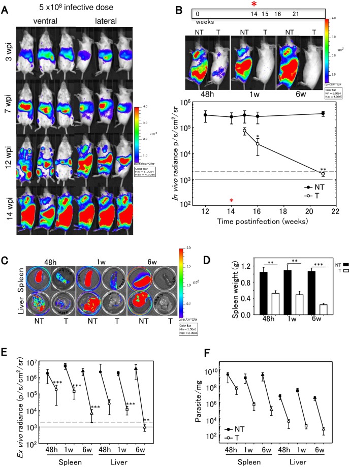
Fig 3. Chronic L. infantum infection in space and time.
A) Representative ventral and lateral view images of Balb/c mice taken at sequential time points over the course of 14 weeks after IV infection with 5 x 10⁸ PpyRE9h luciferase-expressing L.infantum metacyclic promastigotes (representative of n = 30). In the images corresponding to 14 wpi only lateral views are shown because most of BLI signal was detected in this position. Heat-maps are on log10 scales indicate intensity of bioluminescence from low (blue) to high (red); the minimum and maximum radiances for the pseudocolour scale are indicated. B) Animals (n = 15) were treated with 40 mg/kg/d miltefosine via oral for 5 days. Treated and untreated animals were photographed, sacrificed and the spleen and liver imaged at 48 h, 1 week and 6 weeks after the end of miltefosine treatment (15, 16 and 21 wpi). Quantification of lateral bioluminescence for mice shown in B. The red asterisk indicates the start of miltefosine treatment. In vivo radiance from untreated (black circle) and treated (white circle) animals is represented. Black asterisks indicate P-values for t-student test (B,D,E). Comparisons between miltefosine treated groups and untreated control groups (*P < 0.05; **P<0.01; ***P<0.001). Grey line indicates detection thresholds determined as the mean (solid line) and mean +2SDs (dashed line) of background luminescence of control uninfected animals. C) Ex vivo imaging (spleen and liver) in untreated and treated animals at 48h, 1w and 6 w after the end of the treatment (BLI signal results from the D-luciferin injected ex vivo). D) Spleen weights from untreated (black) and treated (white) animals at 48h, 1w and 6 w after the end of the treatment. Each point is the mean ± SD, n = 5 per group. E) Ex vivo bioluminescence signal from spleens and livers obtained from untreated (black symbols) and treated animals (white symbols) at 48 h (circle), 1w (square) and 6w (triangle) after the end of miltefosine treatment. F) Parasite burdens in untreated and treated mice determined by limited dilution assay on livers and spleens.

