
Figure 2.
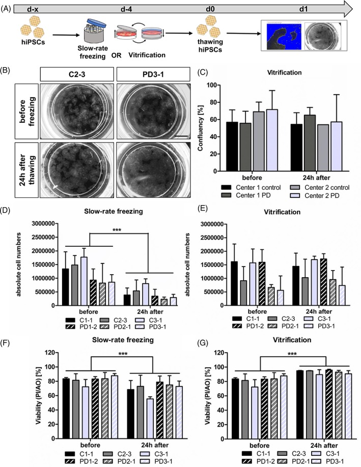
Preservation of confluency, cell numbers, and cell viability by adherent vitrification of hiPSCs. (A): Experimental paradigm. (B): Representative bright‐field images of C and PD hiPSCs in TWIST substrate before and after vitrification. Scale bar 5 mm. (C): Quantification of hiPSC confluency before and after adherent vitrification showed no significant changes. Results are shown as mean ± SD (n = 4 for disease/control group). Statistical analysis: two‐way ANOVA followed by Bonferroni post hoc test. (D–G): Absolute cell numbers and viability of hiPSCs were determined before and after cryopreservation via slow‐rate freezing in suspension (D, F) and adherent vitrification (E, G), as measured by PI and AO staining. Slow‐rate freezing resulted in decreased cell numbers and viability 24 hours after thawing (D). adherent vitrification had no effect on the absolute cell numbers and cell viability (E,G). Results are shown as mean ± SD. Three independent experiments were performed for each hiPSC line (n = 3 per control/disease group). **p value < .005, ***p value < .0001 by two‐way ANOVA followed by Bonferroni post hoc test. see also Supporting Information Figure S1. Abbreviations: AO, acridine orange; C, control; hiPSCs, human‐induced pluripotent stem cells; PD, Parkinson's disease; PI, propidium iodide.