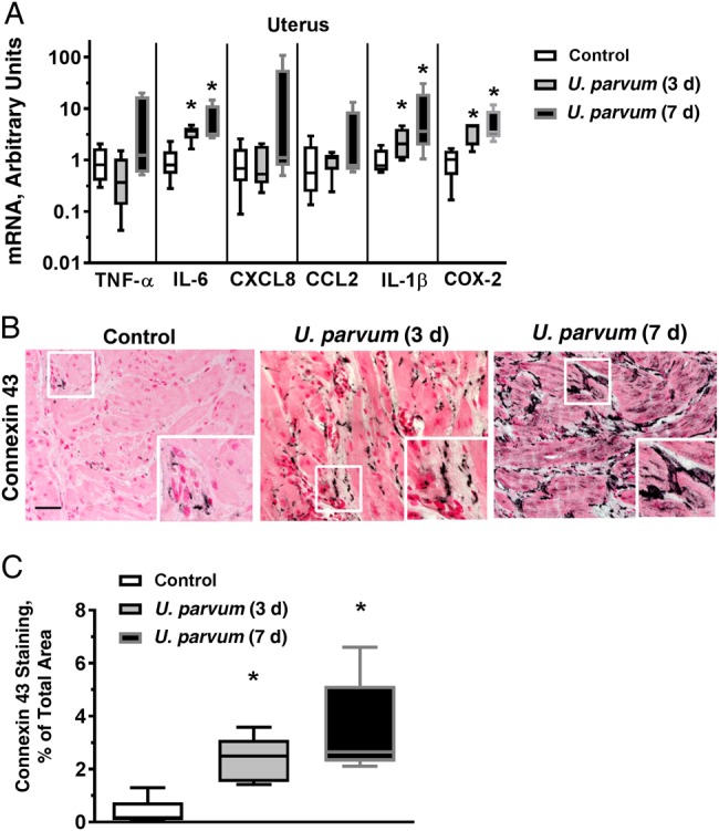
Figure 4.

Intra-amniotic Ureaplasma parvum–induced modest inflammation and increased connexin 43 in the uterus. A, Messenger RNAs (mRNAs) were isolated from full-thickness uterine tissue including the adherent decidua. Quantitative PCR was performed using rhesus-specific Taqman probes. The values were first internally normalized to the endogenous 18S RNA, and the resultant values for the experimental animals were shown as fold increase compared with the mean control value. *P < .05 vs controls (Mann–Whitney U test). B, Immunostaining was performed for the contraction-associated gap junction protein connexin 43. Representative photomicrograph is shown for each study group; the inset in each panel shows a higher-magnification photomicrograph. Note more intensely stained areas of connexin 43 in both U. parvum groups compared with control (scale bar, 50 µm). C, Image quantitation for the connexin 43 immunostaining was performed and expressed as stained area relative to total area (mean of 5 random fields per animal was used as representative value for each animal). Boxes represents 25th–75th percentile; horizontal lines, medians; and whiskers, 5th and 95th percentile values (n = 5 per group). *P < .05 vs controls (Mann–Whitney U test). Abbreviations: IL-1β, interleukin 1β; IL-6, interleukin 6; TNF, tumor necrosis factor.
