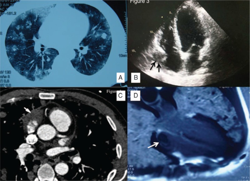
Figure 2.

Radiological presentations of pulmonary metastatic angiosarcoma. (A) Chest computed tomography of pulmonary metastatic angiosarcoma. Multiple unequally sized and sharp-margined nodules are seen, mostly distributed in the peripheral regions. Some ground glass opacities are seen, either surrounding the nodules or unassociated with nodules. (B) Echocardiographic manifestation of cardiac angiosarcoma. A mass is seen arising from the lateral wall of the right atrium (black arrows). (C) Appearance of right atrial angiosarcoma on CT angiography. An irregular soft tissue mass is seen in the right atrium (white arrow). (D) Right atrial angiosarcoma on enhanced cardiac MRI. A low signal was found in the right atrium, but the echocardiographic study was negative (white arrow).
