Figure 2.

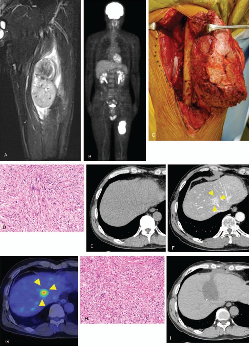
Findings in patient 2. (A) MRI showing a tumor in the left quadriceps femoris muscle, with iso-signal intensity of skeletal muscle on T1-weighted images. (B) FDG-PET scan, showing high accumulation of radioactivity in the tumor of the left thigh and no metastatic lesion. (C) Wide marginal excision of the left thigh. (D) View of the resected specimen, which was pathologically diagnosed as a leiomyosarcoma. (E) CT scan 6 months after wide excision, showing the absence of focal lesion in the liver. (F) Contrast CT scan 3 years later, revealing the presence of focal lesion in the medial liver between the S4 and S8 regions. (G) FDG-PET scan, showing accumulation of radioactivity in the medial liver. (H) View of the resected liver lesion, which was pathologically diagnosed as a hepatic metastasis of leiomyosarcoma. (I) CT scan 1 year after hepatic resection, showing no evidence of local recurrence. CT = computed tomography, MRI = magnetic resonance imaging.
