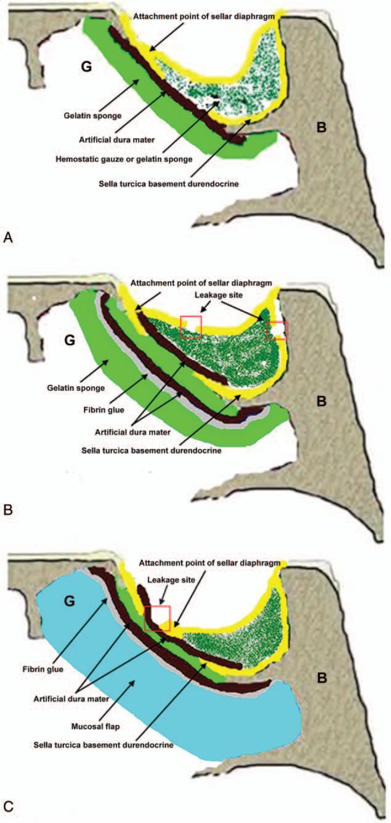
Figure 2.

Schematic diagrams of the 3 surgical methods. (A) Method A. Hemostatic gauze or gelatin sponge was filled in the sellar region. The artificial dura mater was attached outside the sella turcica basement durendocrine. (B) Method B. Hemostatic gauze or gelatin sponge was filled in the sellar region to block the CSF leakage site. The artificial dura mater was patched inside the sellar region. Gelatin sponge was attached at the outside of the sellar region. Fibrin glue was used to achieve fixation. (C) Method C. Hemostatic gauze or gelatin sponge was filled in the CSF leakage site. The artificial dura mater was patched inside the sellar region. The artificial dura mater was also attached outside the sella turcica basement durendocrine. Fibrin glue was used for fixation. A mucosal flap of nasal septum was filled into the defect. B: sphenoid; G: sphenoid sinus cavity. CSF = cerebrospinal fluid.
