
Figure 2.
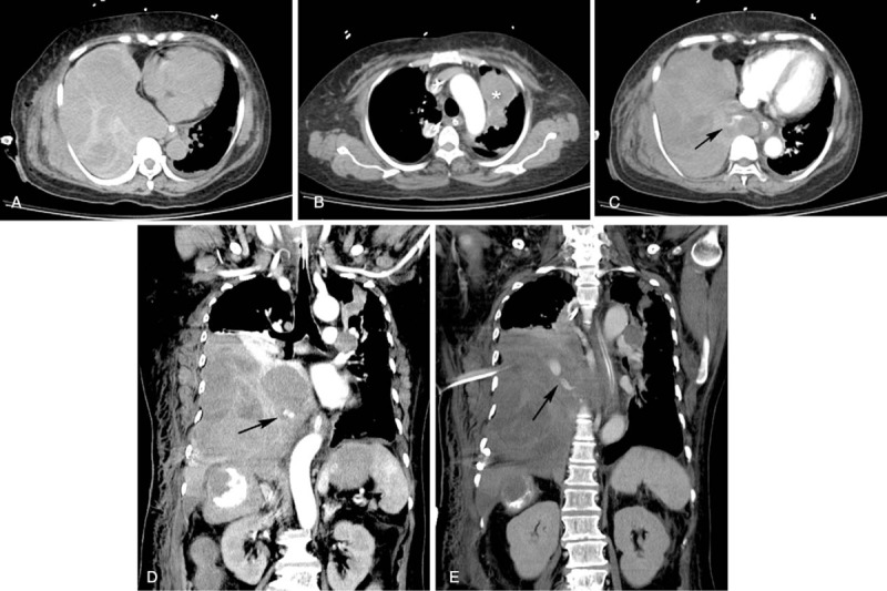
Computed tomography (CT) of the abdomen. (A) Axial noncontrast-enhanced CT revealed a hyperdense lesion in the right pleural space, consistent with hemothorax. (B) Lung metastasis was observed in the left upper lobe of the lung (∗). (C and D) Contrast extravasation was observed on axial and coronal CT angiography (arrows in C and D). (E) Coronal contrast-enhanced CT showed contrast extravasation (arrow).