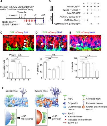
Fig. 6. Neuronal ephrin-B3 and rNSC EphB2 work together to maintain rNSC quiescence.

(A and B) Scheme depicting the experimental procedure of the specifically rescued expression of ephrin-B3 and EphB2 in excitatory neurons and rNSCs in adult EphB2−/−; Efnb3−/− double-knockout mice crossed with the Nestin-CreERT2 mouse line. (C to E) Confocal images showing virus-infected GFP+ rNSCs (arrowheads) in the DG colocalized with EdU (C), rNSCs (arrowheads) with GFAP (D), and neurons (arrow) with NeuN (E). Scale bar, 50 μm. Graphs show the proportion of EdU+ GFP+ rNSCs after EdU treatment (C), GFP+ rNSCs (D), and GFP+ NeuN (E) after 30-day lineage tracing in adult EphB2−/−; Efnb3−/− double-knockout mice crossed with the Nestin-CreERT2 mouse line. In groups a, b, c, d, and e, 3026, 3489, 2887, 3164, and 3165 GFP+ cells of 54, 54, 56, 57, and 56 brain slices were counted, respectively; n = 7 mice for each group. (F) Model for hippocampal quiescent NSCs activated by contacting excitatory neurons through direct neuron-rNSC interaction. In running mice, excited glutamatergic neurons directly interact with rNSCs, which leads to rNSC activation and thereby neurogenesis. This interaction is mediated by ephrin-B3–EphB2 signaling. Results are presented as means ± SEM. *P < 0.05; **P < 0.01; ***P < 0.001; ****P < 0.0001; n.s., not significant.
