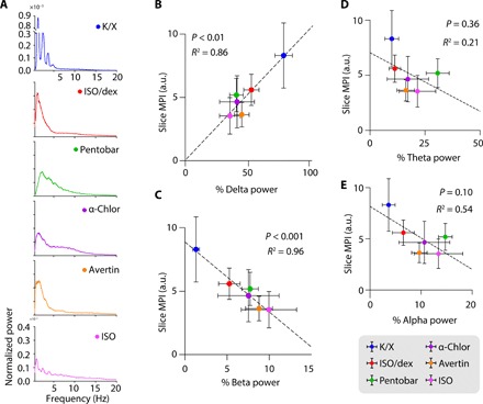
Fig. 3. Glymphatic tracer influx correlates with the prevalence of slow delta waves.

(A) Representative normalized EEG power spectra for each anesthetic regimen. (B to E) Scatterplots depicting the correlation between MPI in coronal slices and the prevalence of delta (B), beta (C), theta (D), and alpha (E) EEG band power. Each dot represents the group mean (whiskers, SD). Correlations were calculated using group means; P values and R2 values are displayed for each correlation. K/X, n = 36 animals for influx and 8 animals for EEG; ISO supplemented with dex, n = 14 animals for influx and 7 animals for EEG; pentobarbital, n = 27 animals for influx and 8 animals for EEG; α-chloralose, n = 20 animals for influx and 8 animals for EEG; tribromoethanol, n = 27 animals for influx and 5 animals for EEG; and ISO, n = 23 animals for influx and 6 animals for EEG.
