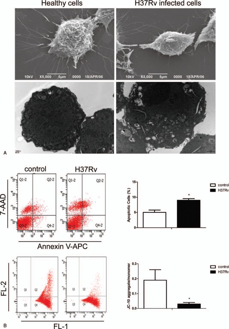
Figure 3.

H37Rv infection promotes RAW264.7 cell apoptosis. Control and H37Rv-infected RAW264.7 macrophages were collected, and SEM and TEM were used to detect cell morphology (A). The apoptosis rate and mitochondrial membrane potential were analysed using flow cytometry (B). Data are expressed as mean±SD from 3 independent experiments. ∗P < .05 versus control. SEM = scanning electron microscope, TEM = transmission electron microscopy.
