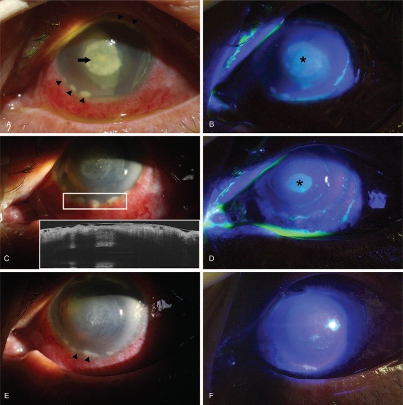
Figure 1.

(A) Slit-lamp photograph of initial presentation. Central, well-demarcated mucopurulent keratitis was apparent (arrow). Well-demarcated round infiltrates were noted at the 6-to-8-o’clock position (arrow heads). (B) Fluorescein staining revealed an elliptical epithelial defect of 4.0 × 4.5 mm in size (asterisk). (C) Five days after admission, considerable improvement in the central mucopurulent lesion had occurred, although aggravation of the corneolimbal infiltrates was noted, along with additional infiltrates at the 6- and 5-o’clock positions (white inlet). Anterior segment optical coherence tomography revealed elevation of the round infiltrates. (D) Fluorescein staining revealed a decrease in the size of the epithelial defect (asterisk), although corneolimbal infiltrates overlying the epithelium remained intact. (E) On the day following initiation of treatment with 1% prednisone acetate eyedrops (arrow heads), considerable improvement in corneolimbal infiltrates was observed. (F) Fluorescein staining revealed complete healing of the epithelium at the site previously affected by P aeruginosa keratitis.
