
Figure 1.
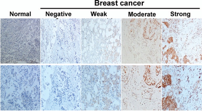
Immunohistochemical analysis of TM4SF1 in breast cancer tissues and normal breast tissues. TM4SF1 showed membranous and cytoplasmic staining. Representative images of sections with different immunostaining intensity. Upper panels: 100×; lower panels: 200×.