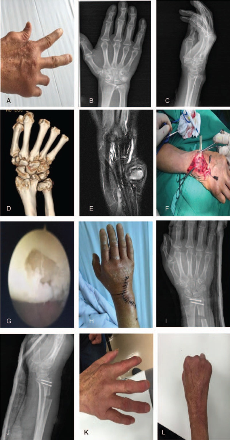
Figure 2.

Case 2: A 79-year-old woman hospitalized due to inability to extend the right little finger since 1 year. Diagnosis: old distal ulnar joint dislocation with osteoarthritis and spontaneous rupture of right ring finger extensor tendon. Distal ulnar articular cleft and TFCC articular cartilage repair was performed assisted by wrist arthroscopy. Sauve–Kapandji osteotomy was performed to treat ulnar joint dislocation, stabilize the ulnar joint; spontaneous fracture of the extensor tendon was repaired. (A–E) General view, X-ray, CT, and MRI images of wrist before surgery; (F) intraoperative view of the broken tendon; (G) wrist arthroscopy revealed intra-articular synovial hyperplasia; (H) general view of patient's hand immediately after the surgery; (I, J) front and lateral X-ray of wrist immediately after the surgery; (K, L) patient's wrist and finger activity, 6 months after surgery.
