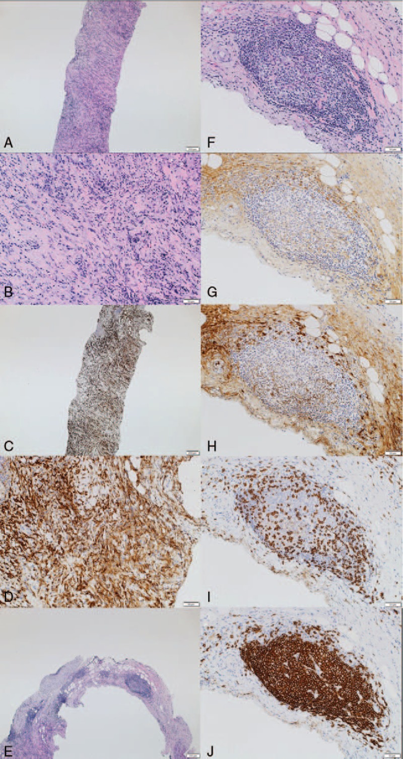
Figure 2.

Histological findings of paravertebral mass in case 1. A: Hematoxylin and eosin staining, low power field; dense lymphoplasmacytic infiltration, and storiform fibrosis were observed. B: Hematoxylin and eosin staining, high power field. C: CD163 staining, low power field; massive infiltration of CD163+ M2 macrophages were observed. D: CD163 staining, high power field. E: Hematoxylin and eosin staining, low power field; hyperplastic ectopic germinal center formation was observed. F: Hematoxylin and eosin staining, high power field. G: IgG staining. H: IgG4 staining. I: CD3 staining. J: CD20 staining.
