Figure 2.

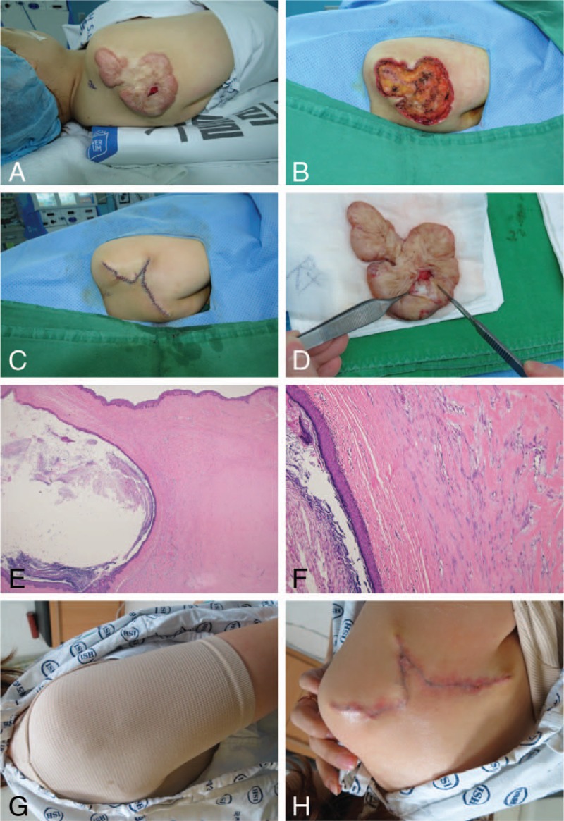
A 43-year-old woman presented with an epidermal cyst arising from a keloid scar and underwent surgical excision (Case 4). (A) The keloid with a centrally located ruptured cystic lesion on the right shoulder. (B) Total excision of the keloid tissue, including the cystic lesion. (C) Immediate postoperative image. (D) Keratinaceous material within the cyst. (E) Histopathologically, a multilayered keratin-filled cyst (left-side) and a dense collagenous keloid (right-side) are present in the dermis in a low-power view. The cyst wall comprised a stratified squamous epithelium with a granular layer (hematoxylin and eosin ×12.5). (F) A high-powered histopathologic view shows that the adjacent dermis contained characteristic broad, eosinophilic, and homogeneous keloidal collagen bundles (hematoxylin and eosin ×100). (G) The patient applies a personalized compression garment for 5 months. (H) Image obtained at 3 months postoperatively.
