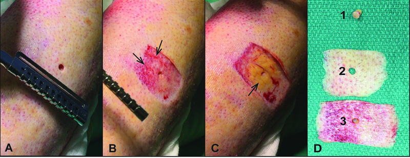
Figure 1 – Intraoperative videography illustrates surgical decision making for excision of burn injured tissue.

Representative patient samples of intraoperative video clips (A-C) of tangential excision (TE) under tourniquet of burned extremity. Biopsy and sequential tangential samples of excised burn tissue were maintained in correct orientation and alignment to capture the superficial to deep excision (D). A) Pre-excision biopsy through burn wound clinically assessed as full thickness at the time of surgery. B) Wound bed after the first tangential excision. Note the hemorrhagic tissue with devitalized appearing dermis, which signified incomplete excision of burn wound (arrows). C) Wound bed after final tangential excision showing healthy appearing fat (arrow). D) Tissue samples corresponding to biopsy (1), first tangential excision (2), and final excision (3).
