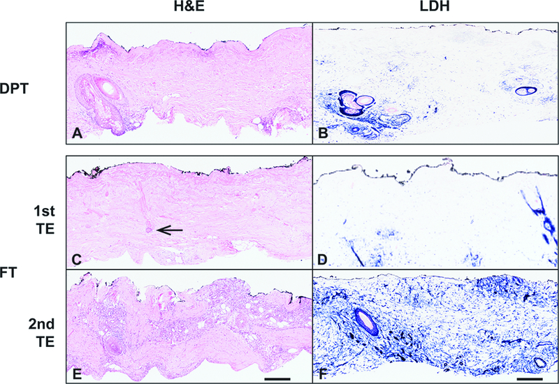
Figure 4 – Viability staining reveals the level of tissue necrosis.

A) H&E staining of a DPT burn. B) LDH staining of same burn wound as in A. C). H&E stain of the 1st TE in FT tissue with necrosis of the upper portions of the excision. The inferior portion of a hair follicle retains aberrant but not clearly necrotic cells (arrow). The 2nd TE contains normal appearing cellularity and skin appendages throughout the tissue. LDH stain sections corresponding to the 1st and 2nd TE reveal viability partially through the 1st TE and throughout the entire 2nd TE. Scale bar = 300 microns. Representative patient samples were chosen.
