
Fig.4. FoxO1 deficiency protects against chronic allergen-induced asthmatic inflammation.
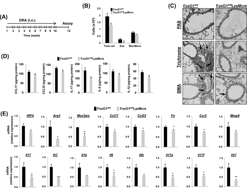
(A) FoxO1fl/fl and FoxO1fl/flLysMcre mice were challenged i.n. with saline or DRA 2 times a week for 10 weeks. End point analysis was performed 2 weeks after the last administration of DRA. (B) The number of BALF inflammatory cells from DRA-challenged FoxO1fl/fl mice were compared with FoxO1fl/flLysMcre mice (N=10). (C) Representative histologic lung sections stained with PAS (black arrowheads), Trichrome (black arrows, peribronchial), and SMA (brown staining) are shown. (D) CCL17, CCL22, IL-17, IL-6, IL-1β cytokines were quantified with ELISA in DRA-exposed lung homogenates (N=6–8). (E) Expression of mRNA for Th2 and Th17 markers in DRA-induced chronic asthmatic lung tissues from FoxO1fl/fl and FoxO1fl/flLysMcre mice. Results are shown as mean ± SE. P values were obtained using a t test. *p<0.05, **p<0.01, ***p<0.001. i.n., intranasal; Eos, eosinophil; Mac/Mono, macrophage/monocyte.